New opportunities for precision medicine
How can we determine whether a drug reaches its intended target in a patient's body? A research team led by the University of Lucerne has developed the first comprehensive framework that links molecular imaging with drug targets, potentially paving the way for more precise therapies.
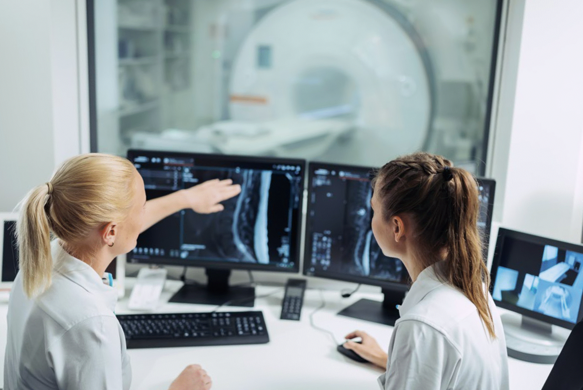
The aim of precision medicine is to ensure that every patient receives the treatment that is most effective for their specific disease. An important part of this approach is the use of drugs that target specific molecules in the body. A key question, however, is whether the corresponding drug target – the molecule to which a drug specifically binds – is actually present in a patient's body. Until now, this has usually been assessed using tissue samples. However, these are invasive and provide only limited information.
This is where molecular imaging comes in. It helps visualize drug targets non-invasively – without damaging tissue or inserting instruments into the body – throughout the entire body and to be monitored over the course of treatment. One example is positron emission tomography (PET). This technique uses so-called tracers, weakly radioactive substances that selectively bind to specific molecules in the body, making them visible on medical images.
New applications for existing imaging methods
To address this challenge, the research team integrated information on more than 1,600 approved drugs, over 700 therapeutic targets, more than 4,000 molecular imaging tracers and gene activity data from over 240,000 patient samples. The resulting framework provides, for the first time, a comprehensive overview of existing imaging methods that could potentially be used to select and monitor drug therapies. The analyses showed that around half of all therapeutic targets can already be visualized using existing imaging techniques. For many of the remaining targets, the researchers identified closely related molecules that can already be detected with available imaging methods. The findings suggest that many existing imaging approaches could also be applied to new drugs and diseases without the need to develop entirely new imaging methods.
"Our long-term vision is to make molecular imaging an integral part of precision medicine," says Martin Walter, Titular Professor of clinical-medical sciences and Head of Nuclear Medicine Research and Development at Hirslanden Klinik St. Anna. "Rather than relying solely on tissue samples or genetic analyses, imaging can show where therapeutic targets are located throughout the entire body and how they change during treatment."
Artificial intelligence connects large amounts of data
The study is based on the integration of numerous biomedical databases using artificial intelligence and modern bioinformatics.
"Artificial intelligence allowed us to identify connections that would have been almost impossible to discover using conventional methods," says Dr. Xiaoying Xu, postdoctoral researcher at the University of Lucerne. "This creates entirely new opportunities to link drugs, diseases and molecular imaging."
Dr. Vincent Taelman, also a postdoctoral researcher at the University of Lucerne, sees considerable potential for clinical application. "Many molecular imaging tracers already exist but are only used for a limited number of applications," he says. "Our work shows that many of these tracers could also be used to support other therapies in the future, without the need to develop entirely new compounds."
Third publication in research series
The study builds on two previous publications by the research group. The Imageable Genome (2023) demonstrated which genes can be visualized using molecular imaging, while The Theranostic Genome (2024) linked diagnostic imaging with targeted therapies. The current study now extends the use of molecular imaging to the therapeutic targets of already approved drugs.
In the long term, the researchers hope that these findings will contribute to more efficient drug development and more personalized patient care.
The study was conducted by researchers from the University of Lucerne, Hirslanden Klinik St. Anna, the University of Geneva, Geneva University Hospitals and international collaborators.
Xiaoying Xu, Vincent Taelman, Pablo Jané, Eduardo Jané, Rebecca A. Dumont, Yonathan Garama, Francisco Kim, María del Val Gomez, Karim Gariani, Martin A. Walter
Repurposing Molecular Imaging to Map Drug Targets in Vivo
Communications Medicine, Nature Portfolio, 2026
Open Access Article


